File:Human brain right dissected lateral view description.JPG
Human_brain_right_dissected_lateral_view_description.JPG (653 × 413 pixel, dimension del file: 40 KB, tipo MIME: image/jpeg)
Somario
| Descrision |
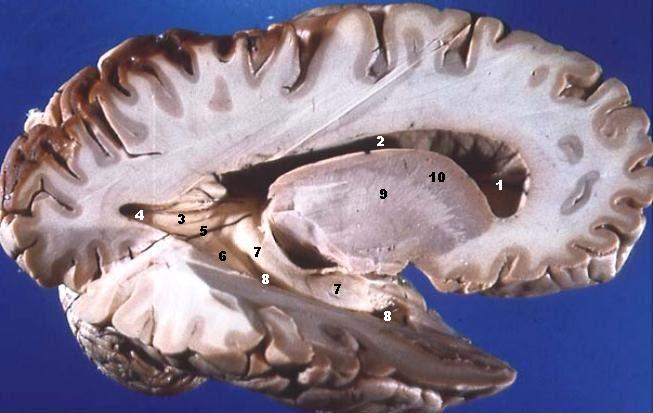
Human brain right dissected lateral view description.JPG Lateral Portion of Frontal, Parietal, Occipital, and Superior Portion of Temporal Lobe Resected. The anterior horn of the lateral ventricle is located in the frontal lobe. The body of the lateral ventricle continues posteriorly into the parietal lobe, the posterior horn into the occipital lobe, and the inferior horn down into the temporal lobe. Some structures produce elevations or bumps in the walls of the posterior and/or inferior horns of the lateral ventricles.
|
| Data | |
| Provenjiensa | http://www.healcentral.org/healapp/showMetadata?metadataId=40566 (Internet Archive of file description page) |
| Autore |
John A Beal, PhD Dep't. of Cellular Biology & Anatomy, Louisiana State University Health Sciences Center Shreveport |
| Liçensa (Ridoparar sto file) |
CC-BY |
| Altre version | http://commons.wikimedia.org/wiki/Image:Human_brain_right_dissected_lateral_view.JPG |
Licensa de doparasion:
- Te si libaro:
- de spartire in jiro – de copiar, distribuir e trasmétar sta opera
- de modifegar – de adatar sta opera
- Soto le seguenti condision:
- atribussion – Te ghe da atribuir ƚa paternità de ƚ'opera ne i modi indicà de l'autor o da chi te gà dà l'opera en lisensa, e en modo tale da no sugerir che lori i sia d'acordo con ti o col modo che te dòpari l'opera.
Il presente file, originariamente pubblicato su https://web.archive.org/web/20110514023714/http://www.healcentral.org/healapp/showMetadata?metadataId=40566, in data 1 novembre 2013 fu esaminato dall’amministratore o dal revisore Avenue, il quale conferma che alla data suddetta l’immagine era disponibile a tale URL sotto la licenza citata.
|
| Annotazioni | Questa immagine è annotata: Vedi le annotazioni su Commons |
Istoria del file
Schicia so on grupo data/or pa vedare el file come che el se presentava in tel momento indegà.
| Data/Ora | Miniadura | Dimension | Utente | Comento | |
|---|---|---|---|---|---|
| In ultima | 16:29, 22 xug 2006 | | 653 × 413 (40 KB) | Patho | {{Information| |Description='''Human brain right dissected lateral view description.JPG''' Lateral Portion of Frontal, Parietal, Occipital, and Superior Portion of Temporal Lobe Resected. The anterior horn of the lateral ventricle is located in the fro |
Doparasion del file
Ła pajina che vien ła dopara sto file:
Doparasion globałe del file
St'altre wiki cua le dopara sto file:
- Uxo de ar.wikipedia.org inte le pàjine
- Uxo de bn.wikipedia.org inte le pàjine
- Uxo de bs.wikipedia.org inte le pàjine
- Uxo de da.wikipedia.org inte le pàjine
- Uxo de de.wikipedia.org inte le pàjine
- Uxo de de.wikibooks.org inte le pàjine
- Uxo de en.wikipedia.org inte le pàjine
- Uxo de en.wikibooks.org inte le pàjine
- Uxo de en.wiktionary.org inte le pàjine
- Uxo de es.wikipedia.org inte le pàjine
- Uxo de eu.wikipedia.org inte le pàjine
- Uxo de fa.wikipedia.org inte le pàjine
- Uxo de fr.wikipedia.org inte le pàjine
- Uxo de fr.wikibooks.org inte le pàjine
- Uxo de gl.wiktionary.org inte le pàjine
- Uxo de he.wikipedia.org inte le pàjine
- Uxo de he.wiktionary.org inte le pàjine
- Uxo de hy.wikipedia.org inte le pàjine
- Uxo de it.wikipedia.org inte le pàjine
- Uxo de ja.wikipedia.org inte le pàjine
Varda ła doparasion globałe de sto file.